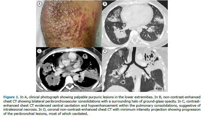
A 42-year-old male smoker presented with a two-month history of fever, hemoptysis, weight loss, and palpable purpura (Figure 1A). Chest CT revealed bilateral peribronchovascular consolidations with a halo of ground-glass opacity (Figure 1B). Follow-up imaging evidenced splenic infarction and progression of the pulmonary lesions, most developing central cavitation (Figures 1C-1D). Laboratory investigation disclosed anemia (10.3 g/dL), leukocytosis (26,000/mm³ with left shift), thrombocytosis (614,000/mm³), glomerulonephritis, complement consumption, elevated C-reactive protein (196 mg/L), and positive c-ANCA antibody; rheumatoid factor, antinuclear antibodies, and tuberculosis testing were negative. Segmentectomy with histopathological analysis confirmed pulmonary granulomatous vasculitis. These findings were compatible with granulomatosis with polyangiitis (GPA). A pulse of high-dose corticosteroid therapy was initiated, but the patient eventually passed away due to massive hemoptysis.

GPA, formerly known as Wegener’s granulomatosis, is a multisystem granulomatous vasculitis that affects small and medium-sized vessels of the upper respiratory tract, lungs, and kidneys.(1) Diagnosis of GPA relies on clinical, laboratory, imaging, and histopathological findings. On imaging, identification of cavitating pulmonary lesions with peribronchovascular distribution and concomitant halo sign, as seen in this case, may help narrowing the differential diagnosis.(2,3) Massive hemoptysis and diffuse alveolar hemorrhage are rare and life-threatening complications of GPA. Treatment usually involves systemic corticosteroids and immunosuppressive agents.
AUTHOR CONTRIBUTIONS RWM was directly involved in reporting the CT scans depicted in this article. RWM, FWL, and DHFL were equally involved in writing, reviewing, conceptualizing, supervising, drafting, and editing the manuscript. Written consent for publication was obtained from the patient.
CONFLICTS OF INTEREST None declared.
REFERENCES 1. Ananthakrishnan L, Sharma N, Kanne JP. Wegener’s granulomatosis in the chest: high-resolution CT findings. AJR Am J Roentgenol. 2009;192(3):676-682. https://doi.org/10.2214/AJR.08.1837
2. Castañer E, Alguersuari A, Gallardo X, Andreu M, Pallardó Y, Mata JM, et al. When to Suspect Pulmonary Vasculitis: Radiologic and Clinical Clues. RadioGraphics 2010;30(1):33-53. https://doi.org/10.1148/rg.301095103
3. Marchiori E, Hochhegger B, Zanetti G. The halo sign. J Bras Pneumol. 2017;43(1):4. https://doi.org/10.1590/S1806-37562016000000354


 English PDF
 Print
Print
 Send this article by email
 How to cite this article
 Submit a comment
 Mendeley
 Pocket